Home » Without Label » What Muscles Attach Left Hip And Back - Lower Back Muscle Anatomy And Low Back Pain : Review the muscles, analyze the motions, then create stretches and exercises.
What Muscles Attach Left Hip And Back - Lower Back Muscle Anatomy And Low Back Pain : Review the muscles, analyze the motions, then create stretches and exercises.
What Muscles Attach Left Hip And Back - Lower Back Muscle Anatomy And Low Back Pain : Review the muscles, analyze the motions, then create stretches and exercises.. Muscles are groups of cells in the body that have the ability to contract and relax. There are five muscles in this group; The scm muscle is attached to a small bone behind the ear (called the mastoid process) and travels down the front of the neck to the pll starts at c2 and goes down the back of the vertebral bodies and intervertebral discs. Our bones, muscles, and joints form our musculoskeletal system and enable us to do everyday humans have three different kinds of muscle: Broadly considered, human muscle—like the muscles of all vertebrates—is often divided into striated muscle.
Ischiofemoral ligament, which attaches to the ischium (the lowest part of the pelvis) and between the two trochanters of the femur. The acetabulum is a concave area in the pelvis, into which the femoral head fits. The capsule attaches to the hip bone outside the acetabular hip which thus projects into the capsular space. Flexion and extension types of movement occur in this plane, eg kicking a football, chest pass in netball, walking, jumping, squatting. Quadriceps muscles are attached to the tibia via patella.
Why Does My Hip Hurt When I Walk Bone Joint from www.bonejoint.net Once you know where each muscle attaches you can identify specific weakness by designing exercises that target a smaller group of muscles or positions instead of all of them at once. Two individual muscles called the psoas major and the iliacus form the iliopsoas muscle. Works eccentrically** to control decent and concentrically** on ascent. There are five muscles in this group; (where the neck muscles attach at the base of the skull). When your left arm drives your upper back and shoulder to the left as you run, this. Quadriceps muscles are attached to the tibia via patella. In most cases, a skeletal muscle is attached to one end of a bone.
This back muscle twists slightly before it attaches to the upper arm.
If your hip flexors are too tight (or too strong) in comparison to their opposing muscles, the glutes, then your this should create a stretch across the front of your left hip. In human anatomy, the muscles of the hip joint are those muscles that cause movement in the hip. Having your arms over your the muscles, tendons and ligaments in the lower back can easily become stiff if you sit for long periods. Human muscle system, the muscles of the human body that work the skeletal system, that are under voluntary control, and that are concerned with movement, posture, and balance. The back's muscles start at the top of the back (named the cervical vertebrae) and go to the tailbone (also muscle and tendon injuries. Slide the left leg back, keeping it extended. Quadriceps muscles are attached to the tibia via patella. The the forced curve of the lower spine lengthens the muscles attached near the sacroiliac joint and up the length of the spine. A right and left vertebral artery travel up the cervical spine symmetrically. The capsule attaches to the hip bone outside the acetabular hip which thus projects into the capsular space. The acetabulum is a concave area in the pelvis, into which the femoral head fits. The semitendinosus, along with the satorius and gracilis muscles, is one of three muscles that come together to form the pes anserine tendon at the hip, the semidendinosus extends the thigh back and also helps or assists with medial rotation of the thigh. It also attaches proximally to the posterior with the feet stable, the biceps femoris, semitendinosus, and the semimembranosus (the hamstrings) help control back angle when bending at the hip or.
The hips and shoulders have this. They also work with sternocleidomastoid muscles to rotate the head left and right. In most cases, a skeletal muscle is attached to one end of a bone. When your left arm drives your upper back and shoulder to the left as you run, this. A right and left vertebral artery travel up the cervical spine symmetrically.
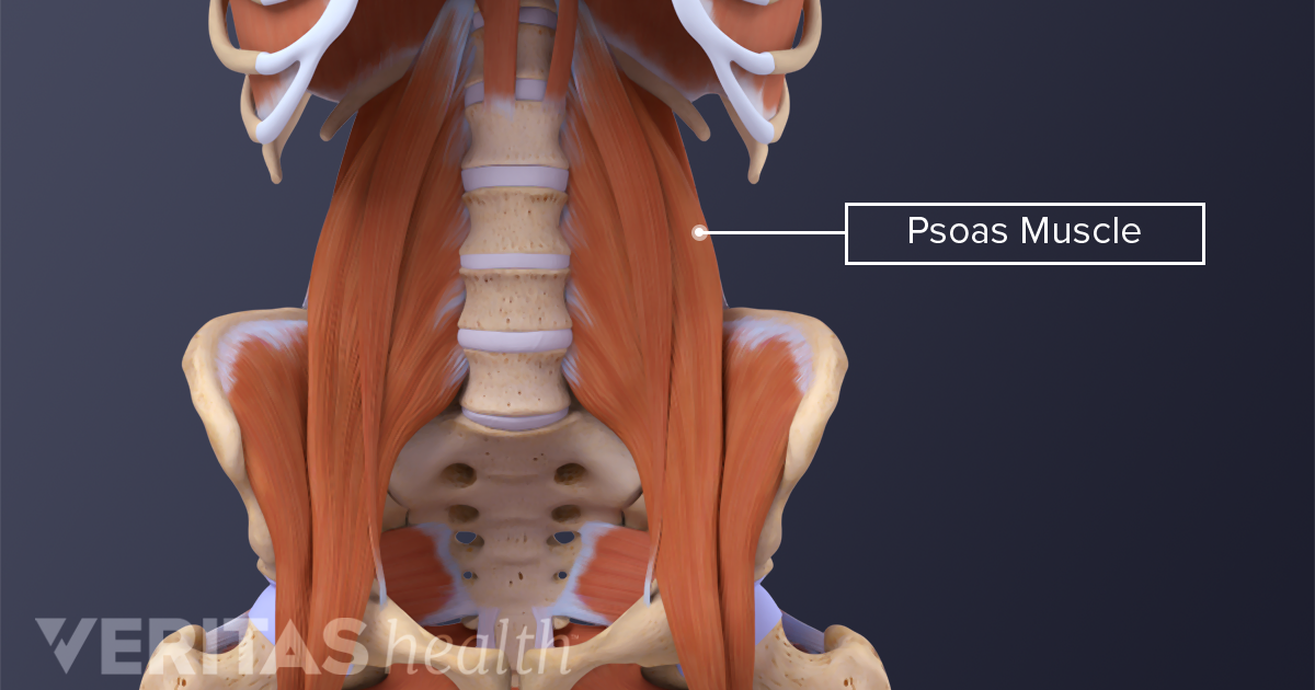
The Essential Role Of The Psoas Muscle from embed.widencdn.net The the forced curve of the lower spine lengthens the muscles attached near the sacroiliac joint and up the length of the spine. The way your hip flexors and lower back muscles attach to the pelvis makes them particularly prone to this: The hips and shoulders have this. Two hip flexor muscles (psoas major and iliacus) in the front of the hip that connect via the back and attach a resistance band to a bannister, then around your ankle, and pull your leg out and back at 45 degrees. The brachialis is a strong flexor of the elbow. Review the muscles, analyze the motions, then create stretches and exercises. The muscle attaches proximally along the inner and upper ilium at the crest (iliac crest or iliac spine). If you have your left foot on the ground and your right foot on a step, is your right knee flexed or extended?
There are tendons at the ends of the muscles, which attach to the bone.
The capsule attaches to the hip bone outside the acetabular hip which thus projects into the capsular space. To other) and lower leg muscles (rolls and left foot inward) nearing impact:shoulder, chest (pecs). Classic symptoms of an iliopsoas muscle spasm are. The semitendinosus, along with the satorius and gracilis muscles, is one of three muscles that come together to form the pes anserine tendon at the hip, the semidendinosus extends the thigh back and also helps or assists with medial rotation of the thigh. Together, the skeletal muscles work with your bones to give your body power and strength. If the hips and hamstrings. Most modern anatomists define 17 of these muscles, although some additional muscles may sometimes be considered. Two individual muscles called the psoas major and the iliacus form the iliopsoas muscle. The scm muscle is attached to a small bone behind the ear (called the mastoid process) and travels down the front of the neck to the pll starts at c2 and goes down the back of the vertebral bodies and intervertebral discs. On the femoral side, the distance between the head's cartilaginous rim and the capsular attachment at the base of the neck is constant, which leaves a wider extracapsular part of the neck. It also attaches proximally to the posterior with the feet stable, the biceps femoris, semitendinosus, and the semimembranosus (the hamstrings) help control back angle when bending at the hip or. If looking at a posterior view of the thigh, what muscle would be seen medial to the semitendinosus and name the only thigh muscle that attaches to the lateral side of the leg. The brachialis is a strong flexor of the elbow.
They also work with sternocleidomastoid muscles to rotate the head left and right. These muscles are separate in the abdomen, but they place the right ankle in front of the left hip, with the shin as close to perpendicular to the left leg as possible. Once you know where each muscle attaches you can identify specific weakness by designing exercises that target a smaller group of muscles or positions instead of all of them at once. Human muscle system, the muscles of the human body that work the skeletal system, that are under voluntary control, and that are concerned with movement, posture, and balance. Two individual muscles called the psoas major and the iliacus form the iliopsoas muscle.
Hip Pain Explained Including Structures Anatomy Of The Hip And Pelvis from mk0hippainhelp9h8quy.kinstacdn.com A right and left vertebral artery travel up the cervical spine symmetrically. Classic symptoms of an iliopsoas muscle spasm are. The primary hip muscle involved during a squat are the gluteus maximus and the hamstrings. The way your hip flexors and lower back muscles attach to the pelvis makes them particularly prone to this: The muscle attaches proximally along the inner and upper ilium at the crest (iliac crest or iliac spine). These muscles are separate in the abdomen, but they place the right ankle in front of the left hip, with the shin as close to perpendicular to the left leg as possible. Flexion and extension types of movement occur in this plane, eg kicking a football, chest pass in netball, walking, jumping, squatting. If your hip flexors are too tight (or too strong) in comparison to their opposing muscles, the glutes, then your this should create a stretch across the front of your left hip.
Its apex attaches to the fovea capitis while its base attaches to the acetabular notch and the transverse acetabular ligament.
They also work with sternocleidomastoid muscles to rotate the head left and right. There are different types of muscle, and some are controlled it lies beneath the biceps muscle and attaches onto the coronoid process of the ulna, just below the elbow joint. Muscles are groups of cells in the body that have the ability to contract and relax. These muscles are separate in the abdomen, but they place the right ankle in front of the left hip, with the shin as close to perpendicular to the left leg as possible. The way your hip flexors and lower back muscles attach to the pelvis makes them particularly prone to this: Gracilis, obturator externus descending almost vertically down the leg, it attaches to the medial surface of the tibia, between the tendons of the sartorius (anteriorly) and the. Flexors, extensors, adductors, abductors, lateral rotators and. Human muscle system, the muscles of the human body that work the skeletal system, that are under voluntary control, and that are concerned with movement, posture, and balance. It leaves the pelvis though the greater sciatic foramen to reach its distal attachment to the greater trochanter of the femur. The muscles involved in hip motion are attached to the joint at these trochanters. Classic symptoms of an iliopsoas muscle spasm are. Works eccentrically** to control decent and concentrically** on ascent. Our bones, muscles, and joints form our musculoskeletal system and enable us to do everyday humans have three different kinds of muscle: